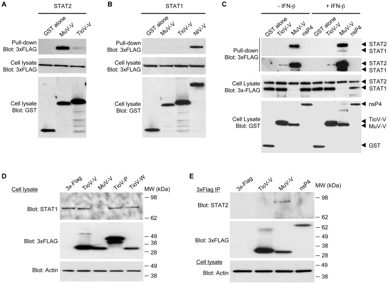
Figure 7. TioV-V fails to interact with human STAT2 and does not induce STAT1 degradation.

(A–B) HEK-293T cells were co-transfected with expression vectors encoding GST alone or fused to MuV-V, TioV-V (A–B) or NiV-V (B) (500 ng/well), and pCI-neo-3xFLAG expression vectors (300 ng/well) encoding for 3xFLAG-tagged human STAT2 (A) or STAT1 (B). Total cell lysates from transfected cells were prepared at 48 h post-transfection (cell lysate; middle and lower panels), and protein complexes were assayed by pull-down using glutathione-sepharose beads (GST pull-down; upper panel). 3xFLAG- and GST-tagged proteins were detected by immunoblotting. (C) HEK-293T cells were co-transfected with expression vectors encoding GST alone or fused to TioV-V, MuV-V or CHIKV-nsP4 (500 ng/well), and pCI-neo-3xFLAG expression vectors encoding for 3xFLAG-tagged human STAT1 and STAT2 (150 ng/well of each vector). At 24 h post-transfection, cells were left untreated or stimulated with recombinant IFN-β at 200 IU/ml. Total cell lysates from transfected cells were prepared at 48 h post-transfection (cell lysate; middle and lower panels), and protein complexes were assayed by pull-down using glutathione-sepharose beads (GST pull-down; upper panels). 3xFLAG- and GST-tagged proteins were detected by immunoblotting. Upper and lower panels on top of figure C correspond to short and longer exposures of the same blot, respectively. (D) HEK-293T cells were transfected with pCI-neo-3xFLAG expression vector (1 µg/well) either empty or encoding for 3xFLAG-tagged TioV-V, MuV-V, TioV-P or TioV-W. Total cell lysates were prepared at 48 h post-transfection and endogenous STAT1 expression levels were determined by western-blot analysis. Actin expression was determined and used as a protein extraction and loading control. (E) HEK-293T cells were transfected with pCI-neo-3xFLAG expression vector (1 µg/well) either empty or encoding for 3xFLAG-tagged TioV-V, MuV-V or CHIKV-nsP4. Total cell lysates were prepared at 48 h post-transfection, and 3xFLAG-tagged viral proteins were purified using anti-FLAG antibodies conjugated to sepharose beads. Co-immunopurification of endogenous STAT2 with 3xFLAG-tagged viral proteins was determined by western-blot analysis (top and middle panel, respectively). Actin expression was determined prior to the immunoprecipitation on total cell lysates and used as a protein extraction control (lower panel).
