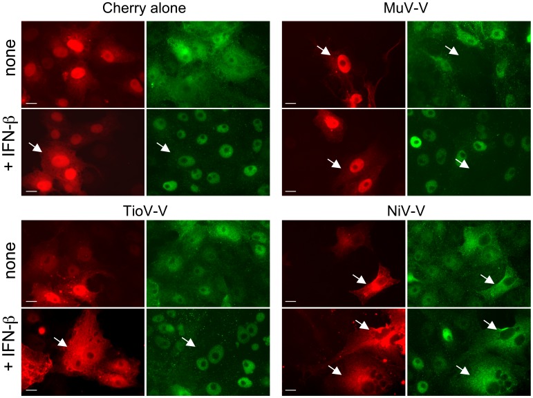
Figure 8. TioV-V does not inhibit STAT1 nuclear translocation induced by IFN-β.

Vero cells were transfected with 100 ng of each plasmid encoding Cherry alone or fused to TioV-V, MuV-V or NiV-V. After 48 h of culture, cells were stimulated with IFN-β for 30 min, and STAT1 was labeled by immunostaining to determine its subcellular localization pattern. Green color corresponds to STAT1 whereas red corresponds to Cherry alone or Cherry-tagged viral proteins. Data show representative fields for each culture condition, and white arrows indicate cells expressing Cherry or Cherry-tagged viral proteins. Scale bar = 10 µm.
