
Figure 5. Examples of middle ear cavities of extinct testudines.
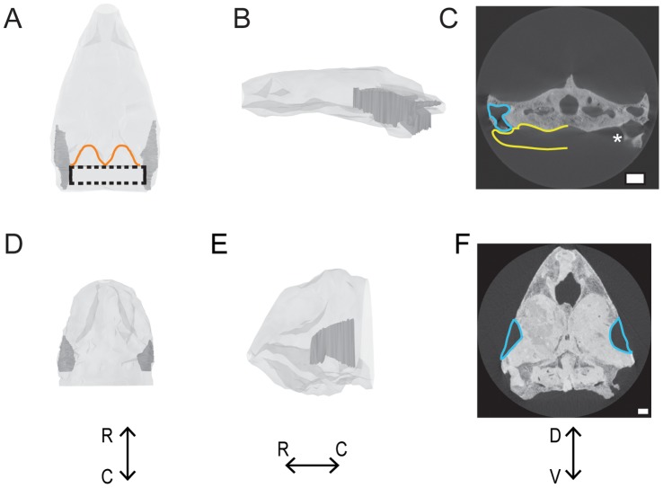
A-C: Connected ears of Galianemys emringeri. Connected middle ears are shown in dark gray; the skull is shown in light gray. The maximum space that the connected middle ears could possibly occupy is indicated by the dashed line. The dorsocaudal edge of the skull is outlined in orange. D-F: Separated ears of Nicholsemys baieri. Isolated middle ears are show in dark gray; skull is shown in light gray. A & D: Dorsal view. B & E: left lateral view. C & F: Transverse view from CT. Middle ear cavities are outlined in blue, and possible extent of middle ear cavity into pharynx is yellow. Asterisk indicates most caudal part of the middle ear cavity that can be seen intact before it opens into the pharynx. Scale bars = 1 cm. R = rostral. C = caudal. D = dorsal. V = ventral.