
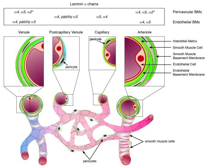
Figure 1. Schematic representation of the cellular and extracellular matrix layers that constitute the vessel wall of arterioles, capillaries, postcapillary venules and venules. Basement membranes underlie the endothelial cell monolayer and ensheath pericytes and smooth muscle cells, and vary in their laminin α chain expression and localization (summarized in the top panel). In arterioles and venules the interstitial matrix interconnects the different cellular and BM layers. *Laminin α2 has not been systematically studied in vascular smooth muscle BMs, but has been reported to occur in smooth muscle of the aorta and carotid arteries.