
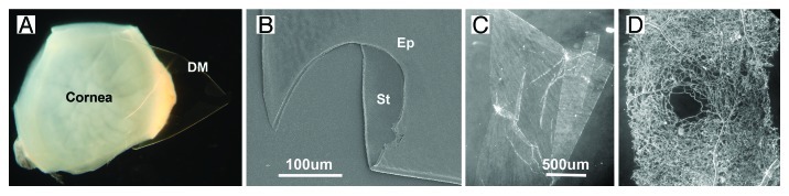
Figure 2. Isolation of BMs and the gross morphology of isolated BMs. The DM is peeled off the inner surface of a detergent-treated human cornea (A). The BM is completely transparent. When imaged by SEM, the DM appears as clean ECM sheet without cellular contamination (B) (St, stromal surface; Ep, epithelial surface). Sheets of isolated ILM and retinal vascular BMs are shown in (C and D). The micrographs show the appearance of the BMs under a dissecting microscope using dark field. The vascular BM sheet (D) was from the foveal area of the retina. Note the foveal avascular zone in the middle of the sample. Bars: (B), 100 µm; (C and D), 500 µm.