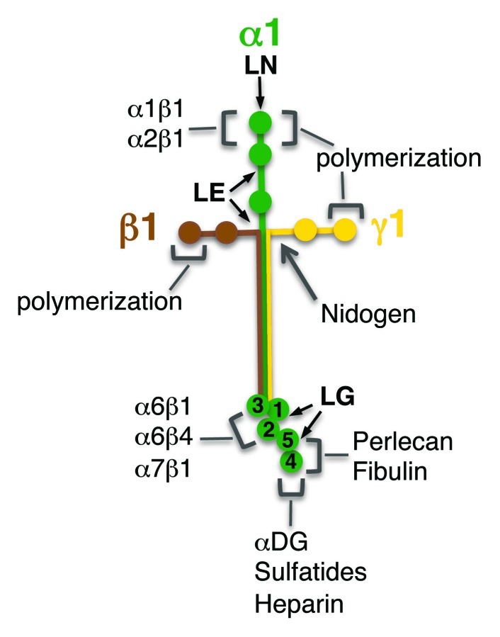
Figure 1. Schematic representation of laminin-111 structure. Each laminin chain is represented with a different color (green, α1; brown, β1; yellow, γ1) and the main interaction domains are indicated (LN, LE, LG). Binding sites for laminin receptors and for other BM components are shown, together with the domains involved in laminin self-assembly. DG, dystroglycan.
