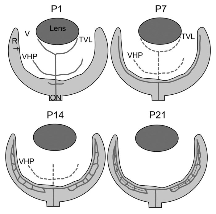
Figure 2. A schematic of retinal vascular development in the normal mouse. Depicted are the key stages in normal vascular development in the mouse. Solid lines indicate blood vessels while broken lines indicate vessels that are regressing. At P1, the hyaloid vasculature in the vitreous (V; white area) consists of the vasa hyloidea proprea (VHP) and the tunica vasculosa lentis (TVL) and the retinal vessels have begun forming. By P7, the superficial vascular plexus in the retina (R) is complete and the TVL and VHP have begun to regress. At P14, the deep retinal plexus is formed and the TVL has regressed. The intermediate plexus forms and the hyaloid vessels completely regressed by P21. The arrow indicates the ILM.
