
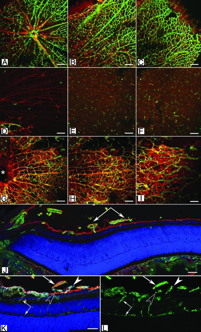
Figure 3 (See opposite page). Retinal vascular defects in the Lama1 mutant mice. (A–I) Retinal flatmounts from P7 mice were stained with GFAP (red; astrocytes) and GS isolectin (green; blood vessels). In the wild type retina (A–C), vessels and astrocytes spread across the entire retina. Hyaloid vessels (arrows) have begun regressing. By contrast, the retina of Lama1nmf223 mice (D–F) has a vastly reduced number of astrocytes with vessels only in the peripapillary region (D). (A, D, G = peripapillary region; B, E, H = midperiphery; C, F, I = far periphery). When the Lama1 mutants retinas are stained with the hyaloid vessels intact (G–I), astrocytes ensheath these vessels and form a membrane in the vitreous. A cross section from a P7 Lama1 mutant stained with GS isolectin (green) and laminin (red) demonstrates the exit of retinal vessels into the vitreous. Cross sections from P10 Lama1nmf223 eyes labeled with PDGFRα (light blue; astrocytes), GS isolectin (green), anti-pan laminin (red; ILM) and DAPI (blue) show vessels from the vitreous branching into the retina. The inner retinal vessels are forming from these diving vessels (paired arrows). Astrocytes expressing laminin (solid arrowhead) on the vitreal side of the ILM (open arrowhead) near the VHP (arrows). Scale bars indicate (A–I), 100 µm; (J–L), 50 µm. Images in this figure were originally published in BMC Developmental Biology.43