
Figure 4. Development of cardiac dysfunction and the death of CMtorKO embryos.
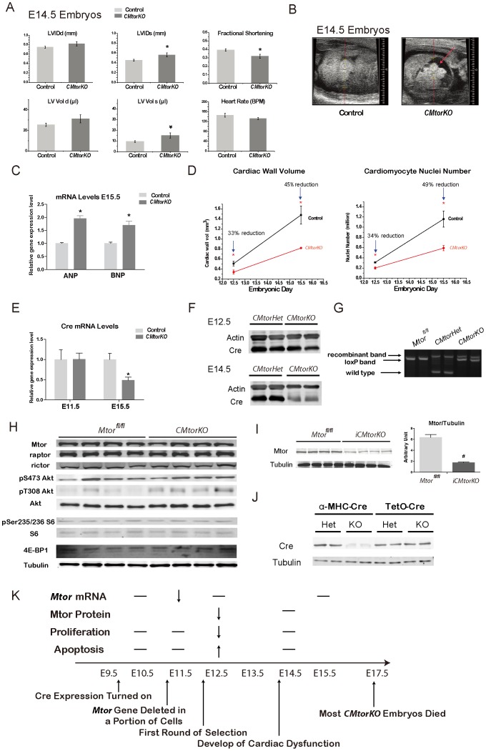
(A) Fetal echocardiography measurements of E14.5 embryonic hearts (n = 9–17). LVIDd: left ventricular interior dimension-diastole; LVIDs: left ventricular interior dimension-systole; LV vol d: left ventricular volume-diastole; LV vol s: left ventricular volume-systole. (B) Representative cardiac echocardiogram of a control embryonic heart (left) and a CMtorKO embryonic heart (right). The arrow indicates pericardial fluid in the CMtorKO embryo. (C) ANP and BNP mRNA levels in E15.5 CMtorKO hearts from live embryos (n = 8). (D) A summary of cardiac wall volume and cardiac nuclei number from E12.5 to E15.5. (E). Cre recombinase transcripts levels in CMtorHet and CMtorKO hearts at E11.5 and E15.5 (n = 8). (F). Western blots of Cre recombinase in E12.5 and E14.5 embryonic hearts. (G). Agarose gel electrophoresis of AC11, AC14 and AC16 PCR products using DNA isolated from E14.5 Mtorfl/fl hearts, CMtorHet hearts and CMtorKO hearts. (H). Western blots of Mtor, raptor, rictor and Mtor downstream signaling molecules in 6–9 week old (adult) failing CMtorKO hearts. (I). Western blot of Mtor protein from adult, doxycycline-induced Mtor deficient hearts (iCMtorKO) (left) and densitometric quantification (right) (n = 4–6). (J). Western blot of Cre recombinase protein from 8-week old CMtorHet, CMtorKO hearts (α-MHC-Cre) and 10-week old iCMtorHet, iCMtorKO hearts (TetO-Cre). (K). A summary of cellular and physiological events in CMtorKO embryos and suggested model of how artificial selection by expressing α-MHC-Cre in mouse heart leads to embryonic lethality. “__“ indicates no change, blank means not measured at the time point. *: p≤0.05 vs. control, #: p≤0.01 vs. control.