
Figure 2. An increase in the mature TACE in Tace-Tg mEFs does not significantly affect the shedding properties of TGFα.
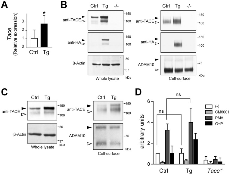
(A) Quantitative RT-PCR analysis of Tace expression in the control (Ctrl) and Tace-Tg (Tg)-derived mEFs. (B, C) Cell surface molecules in mEFs (B) derived from control (Ctrl), Tace-Tg (Tg), and Tace−/− (−/−) mice, and splenocytes (C) derived from control (Ctrl) and Tace-Tg (Tg) were biotin-labeled and analyzed by Western blotting before (Whole lysate) and after (Cell-surface) affinity precipitation with neutravidin beads. The membranes were reprobed with anti-β-Actin antibody and anti-ADAM10 antibody to serve as loading controls for total protein from the whole cell lysate and the cell-surface protein, respectively. Black arrowheads, pro-form. White arrowheads, mature form. Please note that there are small amount of biotinylated pro-form TACE and ADAM10 in panel (C) that were labeled through leakage of the biotin reagent during the procedure. (D) Evaluation of TGFα shedding in the control (Ctrl), Tace-Tg (Tg), and Tace−/− mEFs by a colorimetric assay. Bars, S.D. *p<0.05. ns, not significant.