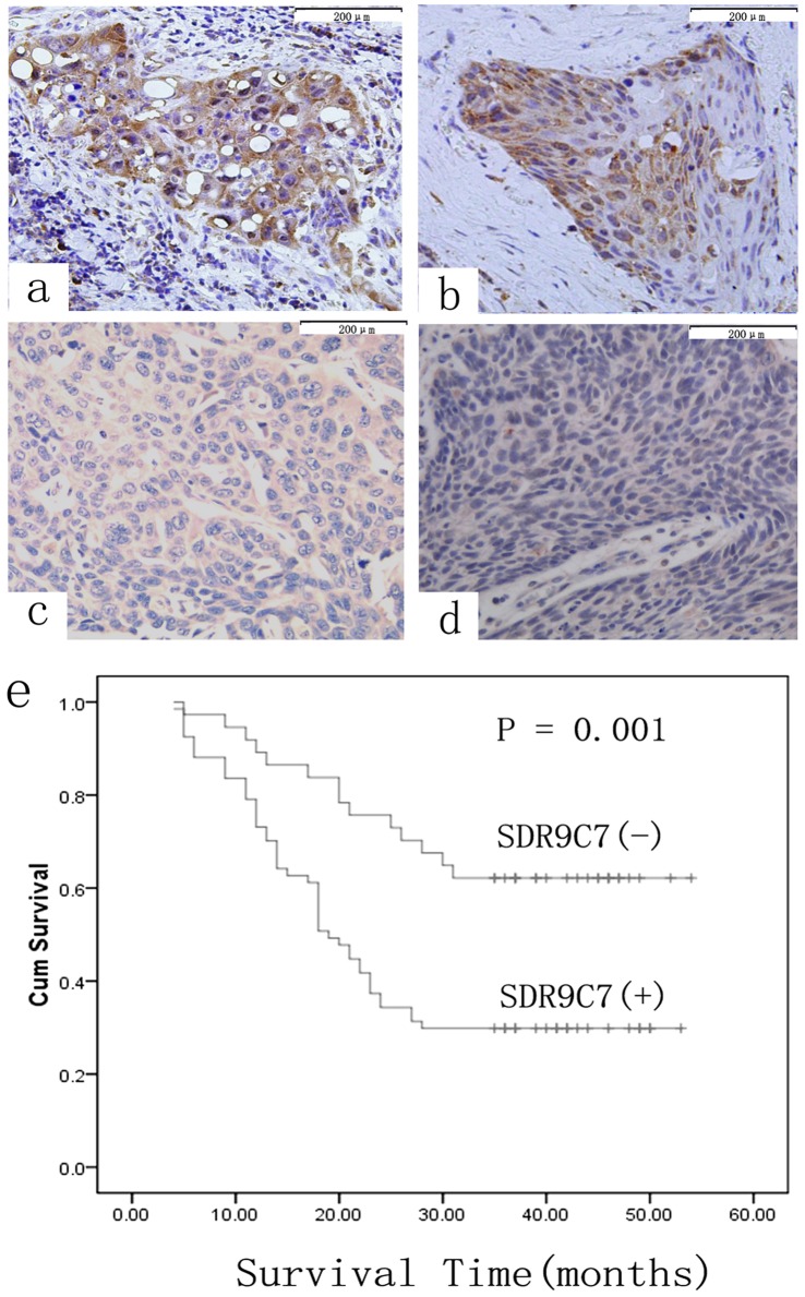
Figure 2. Immunohistochemical staining of SDR9C7 expression in representative ESCC.

The staining of SDR9C7 occurred in cytoplasm of cancer cells. a and b, ESCC tissues with lymph node metastasis; c and d, ESCC tissues without lymph node metastasis original magnification, (SP×200). e, Kaplan–Meier survival curves for patients with ESCC according to the expression of SDR9C7. The survival rate for patientswith positive SDR9C7 expression was significantly lower than that for patients with negative SDR9C7 expression (P = 0.001).
