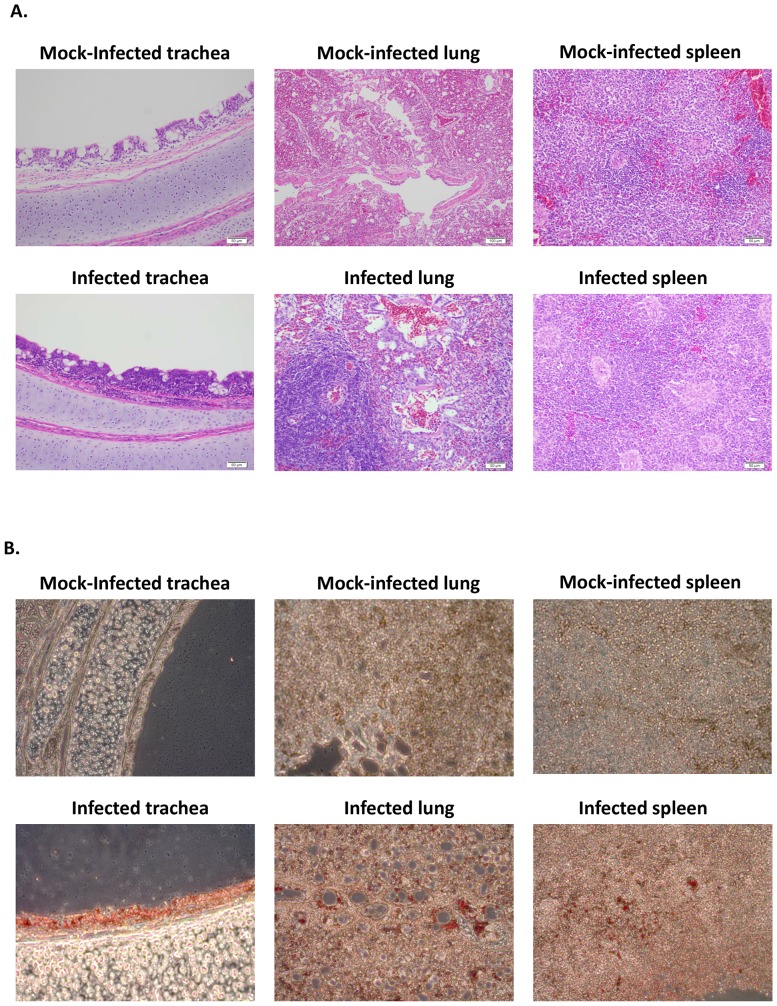
Figure 8. Histopathology and immunohistochemistry in sections of collected tissues from 3-week-old ducks infected with parental and F protein cleavage site mutant APMV-4 viruses.

As described in Table 3, ducks were inoculated with each virus (256 HA units) by the combined intranasal and intratracheal routes, and tissue samples were harvested on 4 dpi. The tissues were fixed with formalin and sections were prepared and stained with hematoxylin and eosin for histopathology (A) or with antiserum against the N protein of APMV-4 for immunohistochemistry (B). (A) Histopathological examination of tissue samples revealed similar microscopic findings in parent and mutant APMV-4 viruses. This is illustrated with representative virus, rAPMV-4. The trachea showed mild to moderate lymphocytic tracheitis with mild to moderate multifocal mucosal attenuation and reduction of tracheal mucous glands. Lung sections exhibited moderate to marked multifocal, lymphohistiocytic bronchointerstitial pneumonia with mild to moderate perivascular cuffing in the pulmonary interstitium. Spleen sections showed moderate reactive lymphoid hyperplasia characterized by expansion of the white pulp by reactive lymphocytes and increase size and cell density of lymphoid follicles. (B) The presence of antigen (stained red) was detected for parental and mutant APMV-4 in the epithelial lining of trachea, in the epithelium surrounding the medium and small bronchi of the lungs and in the spleen.
