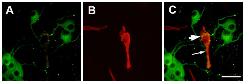
Figure 2. Double fluorescent labeling of MAP-2 (for neurons) and muscle actin (for muscle cells).

Panel A : MAP-2 for DRG neurons; Panel B: muscle actin for SKM cells; Panel C: overlay of Panel A and B. The migrating neurons send axons cross over (thick arrow) and terminate on (thin arrow) the surface of SKM cells. Scale bar = 50 µm.
