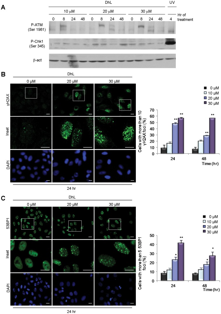
Figure 3. DhL treatment causes the accumulation of DNA damage markers.

Unsynchronized HeLa cells were treated with 0, 20, or 30 µM DhL and lysed or fixed at the indicated times. (A) Immunoblot analysis of phospho-ATM (p-ATM) and phospho-Chk1 (p-Chk1). Representative assay of 3 independent experiments. β-actin was employed as a loading control. Cells were stained with DAPI to visualize the nuclei and treated with specific antibodies for γH2AX (B) and 53BP1 (C). Left: representative fields from 24 h treatment. Insets: magnification of the areas indicated by boxes in the top row. Representative fields for 48 h treatment are shown in Fig. S3. Right: quantification of the number of cells with more than 10 γH2AX foci and more than 5 53BP1 foci. At least 200 nuclei were scored for each sample. Bar: 10 µm. Data represent mean ± SEM of 3 independent experiments. * p≤0.05, ** p≤0.01 vs. control group (0 µM DhL).
