
Figure 5. 20 µM DhL inhibits cell growth by inducing transient arrest in the G2/M phase and permanent accumulation in the G1 phase.
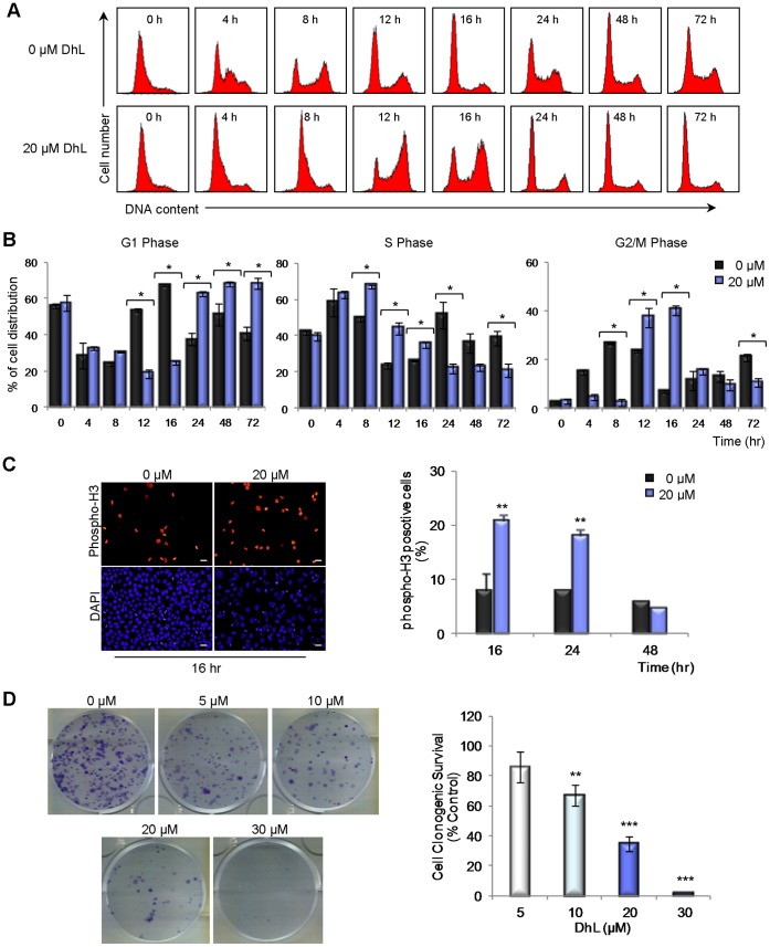
(A) Synchronized HeLa cells were treated with 0–20 µM DhL for 72 h and subjected to DNA flow cytometry at indicated times. Representative DNA distributions from one experiment are shown. (B) Percentage of cells in G1 (left panel), S (middle panel), and G2/M (right panel) phases were determined using the WinMDI 2.9 program. (C) Left: unsynchronized HeLa cells were treated with 0–20 µM DhL and stained with DAPI (to visualize nuclei) and antibodies specific to phospho-H3 at the indicated times. Right: percentage of phospho-H3 positive cells. Bar: 10 µm. (D) Cells were treated with the indicated concentrations of DhL for 48 h, counted, and replated after treatment. Cells that had the ability to form colonies were scored based on clonogenic survival assay 10 days post-treatment. Left: fixed and stained colonies from each treatment representative of 3 independent experiments. Right: number of colonies counted expressed as a percentage of the control (defined as 100%). Data represent mean ± SEM of 3 independent experiments. * p≤0.05, ** p≤0.01, *** p≤0.001 vs. control group (0 µM DhL).