
Figure 3. The MAIT hybridoma response to MR1 mutants.
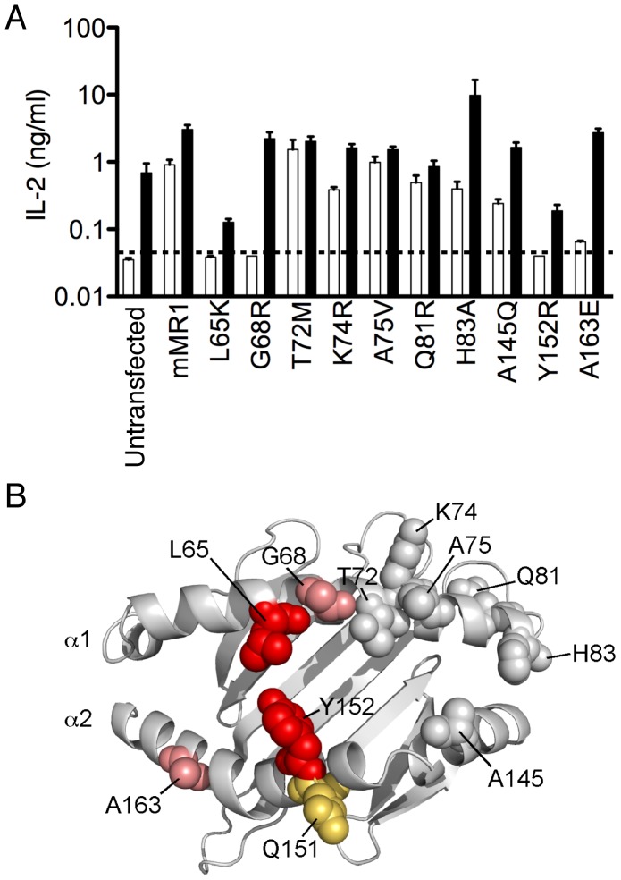
(A) The 6C2 MAIT hybridoma was stimulated with mouse MR1 mutants expressed on LM1.8 fibroblasts in the absence (white bars) or presence of bacteria (black bars) at an MOI of 100. The IL-2 response in both cases could be blocked with an anti-MR1 antibody (data not shown). (B) The effect of the MR1 mutations on both the autoreactive response and the bacterial response is mapped on Phyre-generated mouse MR1 model. Mutations with an effect on both the autoreactive and bacterial response are shown in red, while mutations that only affected the autoreactive response are shown in pink. The residue attributed to the inability of human MR1 to activate mouse MAIT [19] cells is shown in orange.