
Fig. 5.
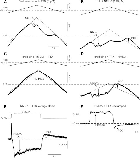
Currents responsible for the NMDA-induced bursting. A: Ca persistent inward current (PIC) in a rat motoneuron recorded in presence of TTX (1 μM), activated by slowly increasing the membrane potential under voltage clamp (top trace), and quantified at its initial peak, where it produced a downward deflection in the recorded current (at arrow) relative to the leak current (thin line). B: voltage-clamp recording of a rat motoneuron in presence of NMDA (100 μM) and TTX (1 μM). Same organization as A. In these conditions, the Ca PIC is hidden by a larger NMDA PIC (downward arrow), and, at the end of the voltage ramp, a persistent outward current (POC; upward arrow) becomes visible. C: voltage-clamp recording of a rat motoneuron in presence of TTX (1 μM) and isradipine (15 μM). Same organization as A. Isradipine is used to eliminate Ca PIC and NMDA PIC interactions by blocking the Ca PIC. As a consequence, the voltage ramp gave a linear current response. D: voltage-clamp recording of a rat motoneuron in presence of TTX (1 μM), isradipine (15 μM), and NMDA (100 μM). Same organization as A. Even when the Ca PIC is blocked by isradipine, clear NMDA PIC and POC are visible on this recording. E: response (bottom thick trace) of a rat motoneuron to a 10-mV voltage step from −77 to −67 mV (top trace). The NMDA PIC is initially visible as a downward deflection in the current trace (downward arrow). The net inward current then decreased continuously after this peak due to the progressive activation of the POC (upward arrow). F: current-clamp recording of the same motoneuron as in E showing the NMDA-mediated depolarization (plateau), which spontaneously turns off and reveals an hyperpolarization phase due to the POC.