
Figure 2. Lysis of IBC cell line, SUM149 by FOXP3-specific T-cells stimulated with FOXP3-encoding RNA-transfected DCs. A.
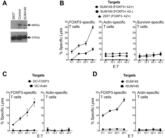
Immunoblot analysis of 48 kDa FOXP3 and 37 kDa control GAPDH protein expression in 293T, SUM149 cell lysates and tonsil tissue lysates. B. Non-adherent PBMCs and DCs were generated from cells obtained from HLA-A2+ healthy donors. DCs were transfected with FOXP3 RNA, survivin RNA and actin RNA and used to stimulate autologous T cells as described in Methods. Post-stimulation the T cells were assayed for lytic activity using a europium-release assay. T cell lytic activity was measured against IBC cells SUM149 and SUM190 and 293T cells as controls. C. DCs expressing FOXP3 and actin were used as targets to demonstrate specificity using effector T cells described in 2A. D. FOXP3-specific T cell cytolytic activity on SUM149 and lapatinib-resistant, rSUM149 cells. Non-adherent PBMCs and DCs were generated from cells obtained from HLA-A2+ healthy donors. DCs were transfected with FOXP3 RNA and actin RNA and used to stimulate autologous T cells as described in Methods. Post-stimulation the T cells were assayed for lytic activity using a europium-release assay. T cell lytic activity was measured against IBC cells SUM149 and rSUM149 cells.