
Figure 3. Immunohistochemistry of phosphorylated Gamma-H2AX in the cortex of an irradiated rat.
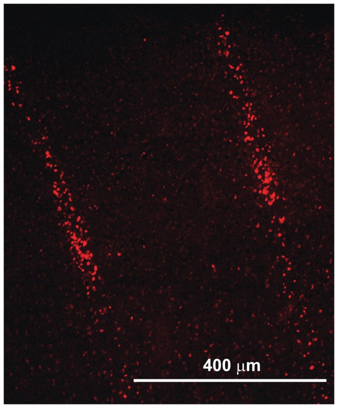
Image shows the microbeam paths across the sensorimotor cortex of healthy rats: apoptotic neurons hit by microbeams (size: 100 µm, c-t-c 400 µm, incident dose 360 Gy) are evident.