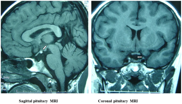
Figure 1. The sagittal (left) and coronal (right) pituitary MRI.

The sagittal image shows the ectopic pituitary (arrow), which is located at the floor of the third ventricle. The coronal image shows absence of a normal pituitary stalk. Also, seen on both images, there is a small anterior pituitary gland.
