
Figure 1. AFM observation of integrase-target DNA complexes.
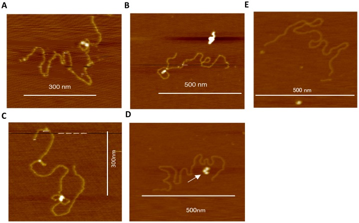
(A, B) Target DNA xkyk bound to HIV-1 integrase forming a loop-like structure or (C) a figure-eight-like structure. (D) DNA showing no HIV-1 integrase binding, where the arrow indicates unbound integrase. (E) Control experiment using x′ky′k including a ScaI digestion site AGTGACT. Bars indicate scale.