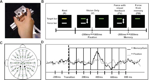
Fig. 1.

A: precision grip apparatus pressed with the subject's thumb and index fingers. B: sequence of conditions for each trial in experiment 1 along with the visual display viewed by the subject. The transition periods that were examined are also shown. C: configuration of electrodes. Clusters of 3 electrodes (green-filled circles) are highlighted with dashed lines showing the 13 regions of interest (ROIs). Black-filled circles are the two reference electrodes used during data collection. D: eight 100-ms time bins used to compare between fixation and memory transitions in experiment 1 and fixation and gain transitions in experiment 2.
