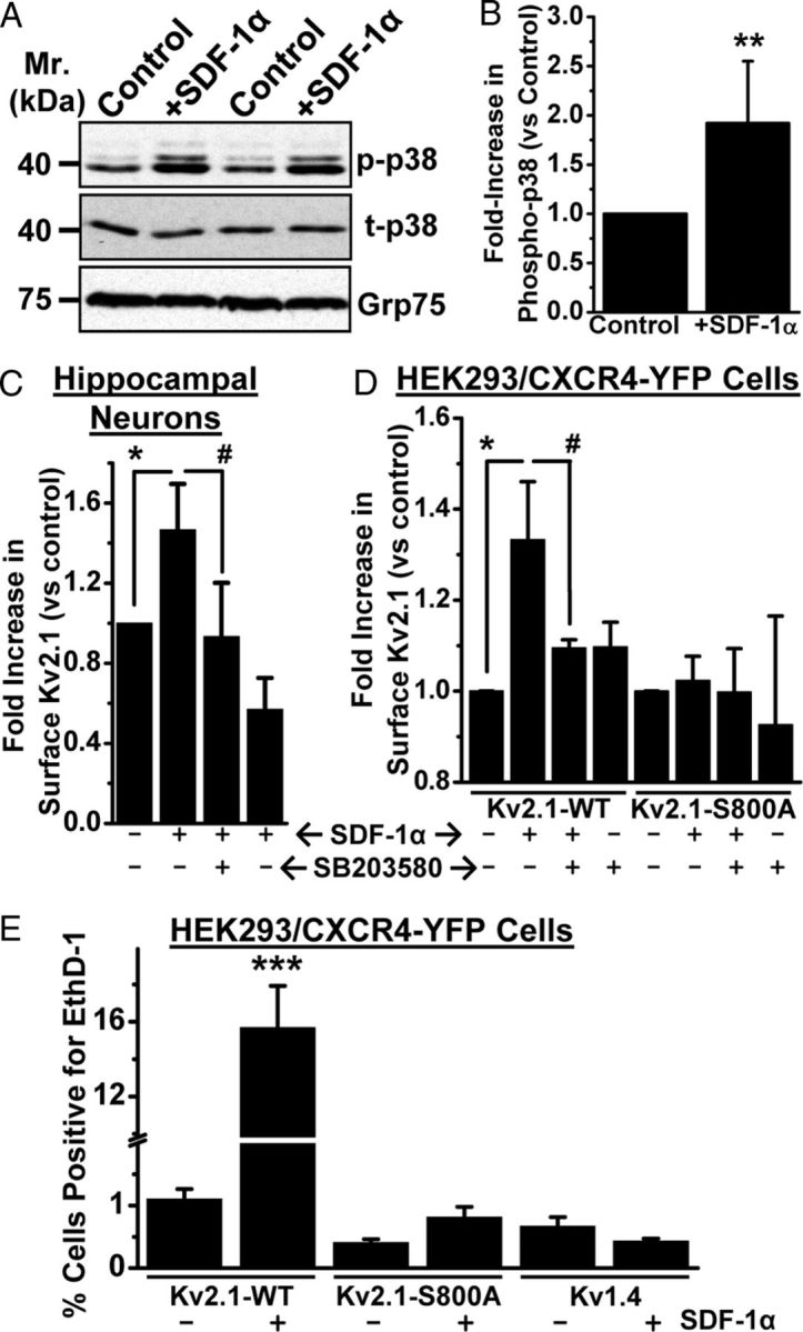
Figure 10.

Sustained SDF-1α treatment leads to increased surface expression of Kv2.1 that is dependent on p38 MAPK phosphorylation at residue S800, which governs the induction of cell death. A, B, Prolonged treatment with SDF-1α (100 nm, 3 h) led to increased phosphorylation of p38 MAPK in cultured rat hippocampal neurons, as determined by immunoblot analysis (A) and quantification by densitometry from three independent experiments (B). **p < 0.01 versus control (Student's t test). C, D, Increase in the level of surface biotinylated Kv2.1 protein, as determined by immunoblot analysis, from cultured rat hippocampal neurons (C) and HEK293 cells expressing Kv2.1–WT and CXCR4–YFP (D) during SDF-1α treatment (100 nm, 3 h), which can be attenuated by pretreatment with SB203580 (1 μm). No increase in the level of surface biotinylated Kv2.1 protein was observed during SDF-1α treatment of HEK293 cells expressing Kv2.1–S800A mutant and CXCR4–YFP (D). For details of biotinylation experiments and densitometry analysis, see Materials and Methods. Data are presented as mean ± SEM from three independent experiments. *p < 0.05 versus untreated conditions; #p < 0.05 versus SDF-1α treatment group (one-way ANOVA with post hoc Bonferroni's correction). E, Increased cell death during sustained SDF-1α treatment (100 nm, 3 h) was absent in HEK293 cells expressing CXCR4–YFP and Kv2.1–S800A mutant, as well as in HEK293 cells expressing another Kv channel, Kv1.4. Data are presented as mean ± SEM of percentage of cells positive for EthD-1 (n = >500 cells from 4 independent experiments). *p < 0.05 and ***p < 0.001 versus untreated conditions; #p < 0.05 versus SDF-1α treatment group (one-way ANOVA with post hoc Bonferroni's correction).
