
Figure 7. Permissiveness of PDA cell lines to VSV-ΔM51-GFP, VVT7 and HSV-1.
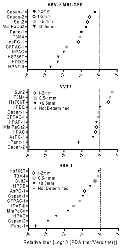
Cells were infected with serial dilutions of virus and, after a 1 h absorption, overlayed with media containing 0.5% BactoAgar. VSV-ΔM51-GFP foci were counted by fluorescent microscopy at 3 d p.i. VVT7 and HSV-1 plaques were counted after staining with crystal violet at 5 d p.i. Titers are expressed relative to those on Vero cells. A relative yield of 0 indicates that the PDA cell line and Vero are equally permissive to the virus, while negative numbers indicate reduced permissiveness on the PDA cell line. Plaque size was determined for all viruses at 5 d p.i. following crystal violet staining.