
Fig. 10.
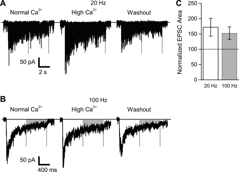
Synaptic transmission during HFS was enhanced by increasing extracellular Ca2+ concentration. EPSCs (n = 8 cells from 8 different slices) were recorded during 20- and 100-Hz stimulation in normal (2.0 mM) and elevated (3.8 mM) extracellular Ca2+ (with Mg2+ concentration adjusted to maintain constant total divalent cation concentration). A: representative example of EPSCs recorded during 8 s of 20-Hz HFS (160 stimuli total); EPSCs were recorded in normal-Ca2+ ACSF, during application of high-Ca2+ ACSF, and after washout in normal-Ca2+ ACSF. Holding current level is indicated by the solid horizontal line; the second half of the HFS (stimuli 81–160) is indicated by the two vertical dashed lines. B: representative example (same cell as shown in A) of EPSCs recorded during 1.6 s of 100-Hz HFS (160 stimuli total). C: data summary of all 8 recordings. Synaptic current areas during the second half of the HFS were summed and normalized relative to the EPSC area recorded in normal-Ca2+ ACSF. For both 20- and 100-Hz HFS, EPSC areas were significantly increased during application of high-Ca2+ ACSF.