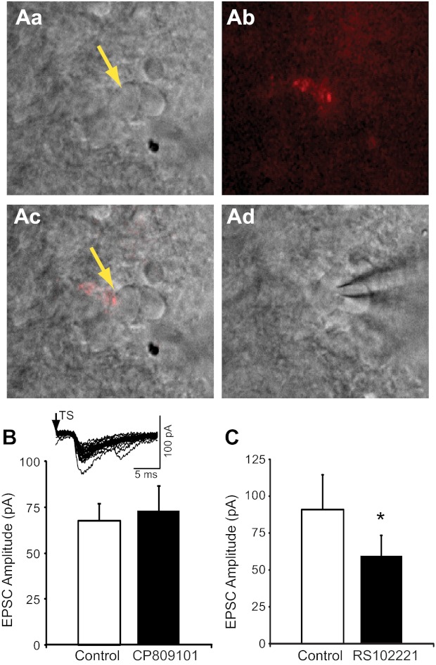
Fig. 9.

5-HT2CRs modulate synaptic transmission from chemosensory afferents in the nTS. A: example of a DiI fluorescent synaptic bouton labeled from the carotid body on a caudal nTS cell. Aa: infrared-differential interface contrast (IR-DIC) image of nTS cells medial to the TS. Yellow arrow denotes labeled cell. Ab: DiI labeling (pseudocolored red) of synaptic boutons from chemoafferent fiber visualized under fluorescence. Ac: overlay of images from Aa and Ab illustrates that the fluorescent boutons were on the soma of the nTS neuron. Ad: after identification of a bouton-labeled nTS cell, a patch electrode was guided to the cell under IR-DIC for recording. B: group raw data (n = 6) of TS-EPSC amplitude during control and CP809101. Inset: representative example of TS-EPSCs evoked from a DiI-labeled nTS cell. Note the low variability of TS-evoked EPSCs, indicative of a monosynaptic cell. C: group raw data (n = 7) of TS-EPSC amplitude that was reduced by RS102221 compared with control. *P < 0.05.
