
Figure 1.
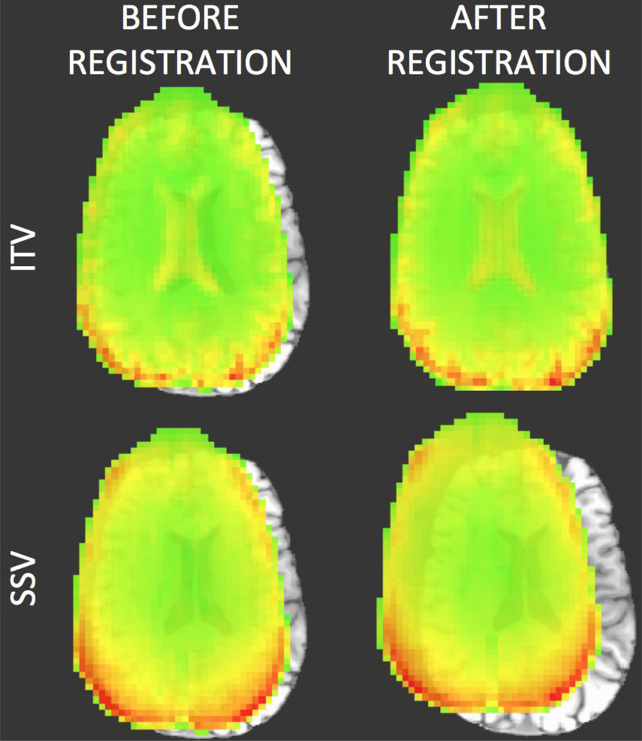
Representative case of anatomical-to-functional alignment in which alignment failed when the EPI SSV was used as the target (bottom row), but did not fail when the ITV was used as a target (top row). In all four cases, the underlay is the MPRAGE volume being aligned towards the EPI data. The overlay on the top row corresponds to the ITV prior and posterior to alignment. In the lower row the overlay corresponds to a SSV prior and posterior to alignment. In addition to the differences in the alignment results, this figure also highlights how the ITV contains higher structural information (e.g., the ventricles are clearly observed) that is not present in the SSV.