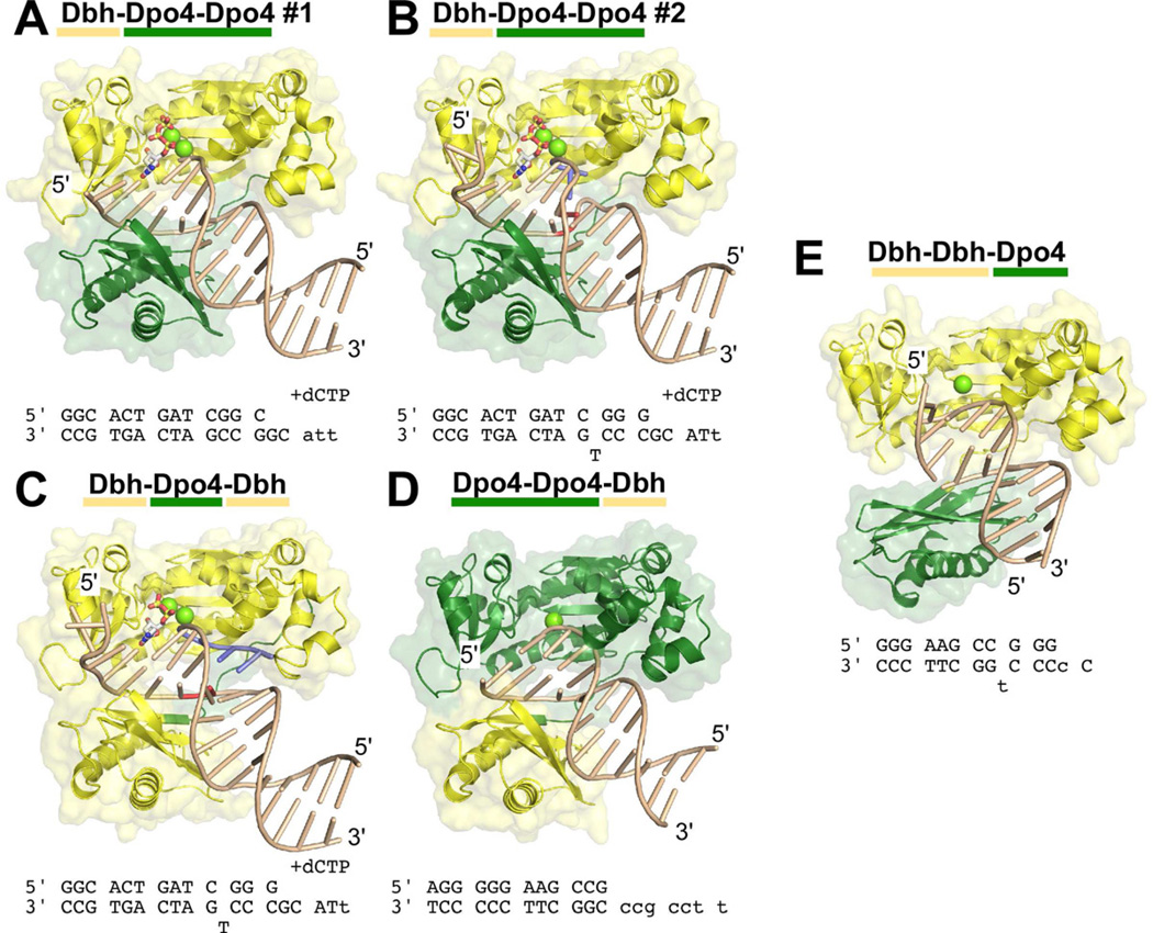
Figure 3. Structural Overview of Chimeric Polymerases.

The structures of five complexes between chimeric polymerases and DNA are shown: (A) Dbh-Dpo4-Dpo4, complex #1, (B) Dbh-Dpo4-Dpo4, complex #2, (C) Dbh-Dpo4-Dbh, (D) Dpo4-Dpo4-Dbh, and (E) Dbh-Dbh-Dpo4. The protein is shown with a molecular surface representation and is colored by the parental sequence: Dpo4 (green), Dbh (yellow). DNA primer-template sequences and nucleotide (if any) that were included during co-crystallization are shown below each structure; sequences shown in lower case were not visible in the electron density maps. DNA is shown in a ladder representation. Nucleotides shown in red (in B and C) were designed to be unpaired; nucleotides shown in blue (in B and C) were added to the primer-terminus during co-crystallization (see Supplemental Material, Figure S3 D and E). Calcium ions at the active site are shown as chartruse spheres; incoming nucleotides are shown in stick representation, colored by atom: carbon (white), oxygen (red), nitrogen (blue), phosphate (yellow). See Figures S3 and S4 for additional details.
