
Figure 1.
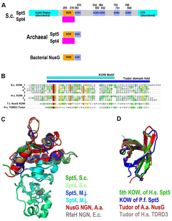
(A) Domain organization of Spt4 and Spt5 proteins. Domain boundaries are drawn to proportions according to their polypeptide lengths. (B) Alignment of sequence segments from Spt5 and NusG proteins that encompass the KOW motif. Note a Tudor domain sequence of TDRD (Tudor domain-containing protein 3) is also included. Color variation (from red to green) reflects degree of conservation (invariant to similar). (C) Structural superposition of homologous Spt4 and Spt5 proteins. Protein IDs are indicated with the different colors. (D) Structural superposition of KOW domains of Spt5 and NusG together the Tudor domain of TDRD3. Abbreviations: S.c. - Saccharomyces_cerevisiae; M.j. - Methanococcus jannaschii; A.a. - Aquifex aeolicus; P.f. - Pyrococcus furiosus; T.t. - Thermus thermophilus; E.c. - Escherichia coli; H.s. - Homo sapiens; and TDRD3 - Tudor domain-containing protein 3.