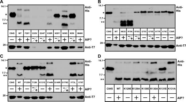
Figure 4.

Peptidase assay results with AgrB point mutants. In each panel, T7-tagged AgrB mutants were expressed along with His6-AgrD in E. coli. Cleavage of His6-AgrD was determined through immunoblots for the His6 tag (upper blots). Lower blots show the presence of T7-AgrB as detected with anti-T7-HRP. Also in the panels, “+” indicates whether any AIP is detectable when the AgrB mutant is expressed in E. coli and S. aureus, “−“ indicates no AIP was made in either. “−/+” indicates that AIP is made in S. aureus but not E. coli. Panel A, AgrB mutations F26Y to M46K; panel B, N63S to C84R; panel C, P125H to I176T; and panel D, K129A/E to K131A/E. For interpretation of the cleavage assay, “*” indicates full-length His6-AgrD, and “**” indicates cleaved His6-AgrD with the C-terminal 14 residues removed.
