
Figure 6.
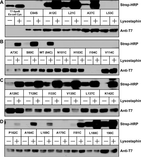
Mapping AgrB topology in S. aureus Δagr using SCAM. In each panel, T7-tagged AgrB (C84S) containing indicated cysteine substitutions were expressed in S. aureus Δagr (AH1292). Lanes marked with a “+” were pretreated with lysostaphin to expose intracellular cysteines prior to labeling. 50 μM MPB was used in untreated and pretreated conditions to label cysteines. Following immunoprecipitation using anti-T7 agarose, elutions were immunoblotted with Strep-HRP for the MPB label (top blots) and anti-T7-HRP to detect T7-AgrB (lower blots). T7-SpsB containing an extracellular cysteine was used as positive control for labeling. Panel A, T7-SpsB to L53C; panel B, A73C to V114C; panel C, A126C to K142C; and panel D, P162C to 190C.