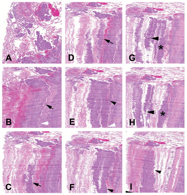
Figure 1.

(A) Top view of 3D-reconstructed image of lung adenocarcinoma showing tumor islands. (B–J) Fifty whole-slide images of a serial-sectioned paraffin embedded specimen were combined and a 3D image was obtained to study the structure of the tumor islands and its relation with surrounding structures. (B–D) Different planes of view are shown depicting the islands running deep into the tissue (arrows). (B, E–J) Neighboring islands tend to connect (arrow heads and asterisks) and at certain points merge with the main solid tumor (white arrow heads).
