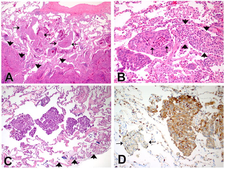
Figure 2.

(A) An example of lung adenocarcinoma with tumor islands. The islands are isolated within airspaces (arrows) and are several alveoli away from the main tumor (arrow heads). (B) A high-power view shows clusters of atypical cells with necrosis (arrows). The arrowheads indicate a collection of benign alveolar macrophages in the adjacent air space with significant difference in cytology between the two groups. (C) Another example of lung adenocarcinoma with tumor islands. The islands are present in the alveoli adjacent to the blue-inked wedge resection margin (arrowheads). (D) Keratin stain highlights the tumor islands confirming an epithelial origin. Conversely, a cluster of alveolar macrophages (arrows) are negative for keratin. (Immuno stain for pan-keratin on a deeper section of C).
