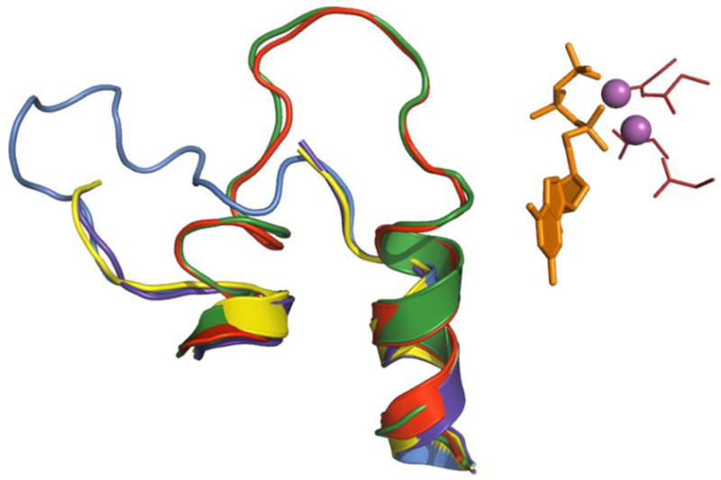
Figure 3. Structural variants of the trigger loop in yeast RNA polymerase II.

Elements of the active site (from GTP-bound 2e2h) are represented as in Fig. 2. (substrate is orange sticks, catalytic Mg2+ cations are solid magenta spheres, catalytic tetrad is red sticks) Superimposed are trigger loops (cartoon) from NTP-free enzyme (1sfo, purple blue), GMPCPP-bound 2e2j (yellow) and 2nvt (marine), and UTP-bound 2nvz (red).
