
Figure 1. Schematic structure of LFn-DT-TGF and expression and cleavage analyses.
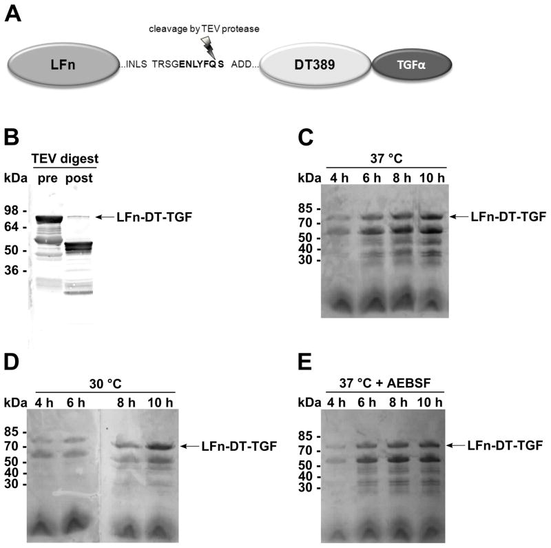
(A) The N-terminal 254 amino acids of LF are fused via a linker containing a TEV protease cleavage site to the 389 N-terminal amino acids of diphtheria toxin and TGFα. (B) TEV protease cleavage of LFn-DT-TGF in the supernatant of B. anthracis BH460 (10-fold concentrated). Immunodetection by anti-diphtheria toxin. (C-E) Expression of LFn-DT-TGF in B. anthracis strain BH460 at 30 or 37 °C and in the presence of 167 μM protease inhibitor AEBSF. Supernatants were collected after 4–10 h, 10-fold concentrated and detected by SDS-PAGE and subsequent Coomassie staining.