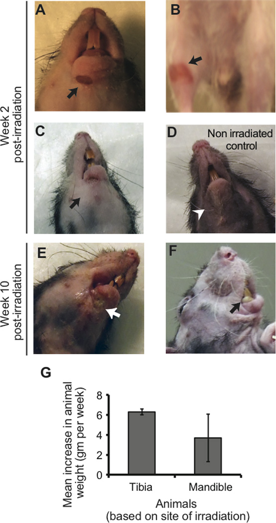
Fig. 2.

Localized radiation outcomes. Mandible (A) and tibia (B) developed localized radiation-induced erythema (black arrows) within the first two weeks post-irradiation. In all animals, associated inflammatory edema was significantly pronounced in the irradiated mandible (C, black arrow) compared to non-irradiated animal (D, white arrow head). Orofacial soft and hard tissues rapidly progressed to necrosis (E, white arrow) and formation of bone sequestrum (F, black arrow) by week 10. Soft tissue overlying irradiated tibia remained intact as shown in B up to week 20. The mean cumulative weight gain of all animals with irradiated mandible was lower and more variable (wide error bars) than those with irradiated tibia [(G) but the differences were not statistically significant].
