Fig. 3.

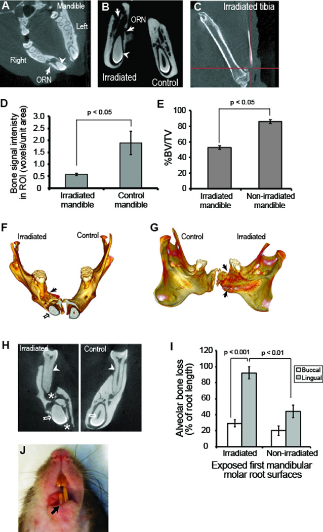
Skeletal and dental outcomes of irradiation. Micro-CT showed osteoradionecrosis (ORN, white arrows) and opacification of incisor tooth (white arrowhead) that developed in irradiated right mandible about 10 weeks post-irradiation Non-irradiated (control) left mandible and teeth retained normal anatomical trabecular pattern and patent pulp chamber respectively (A and B). Tibial cortical bone plate remained intact post-irradiation without any radiological signs of ORN up till time of sacrifice at 20 weeks (C). There was marked reduction in bone quantity in irradiated right mandible relative to non-irradiated control left mandible (P < 0.05) (D, E). Three dimensional micro-CT volume rendering of mandible demonstrated advanced alveolar bone loss (black arrows, F, G). Representative coronal view of mandibular ORN at the level of the first molar (H) displayed severe periodontal bone loss (white arrowheads), trabecular bone loss (white star) and pulpal calcification of both molar and long incisor teeth (white arrow and arrowheads). Periodontal bone loss was notably more severe on the lingual than the buccal side in both irradiated (p < 0.001) and non-irradiated mandible (p < 0.01) (I). There was also delayed eruption of right mandibular incisor relative to the left (J).
