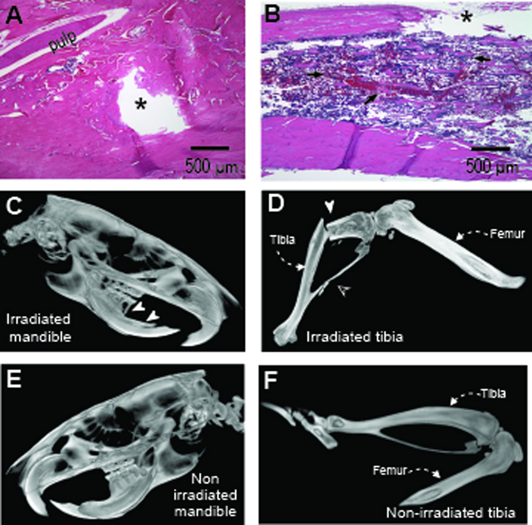
Fig. 5.

Trauma complicates osteoradionecrosis. Hematoxylin and eosin histological sections show trauma-induced cortical window (black star) in irradiated mandible (A) and tibia (B). The irradiated mandible displayed marked acellularity, micro-fractures and pulpal atrophy while tibia displayed disorganized bone trabeculae and adipocytic marrow infiltrates (black arrowheads). Micro-CT volume rendering of irradiated (C and D) and non-irradiated sites (E and F) showed advanced alveolar and periodontal bone loss in irradiated mandible (white arrowheads) relative to non-irradiated site; irradiated tibia with associated fibula succumbed to fracture (white and clear arrowheads respectively).
