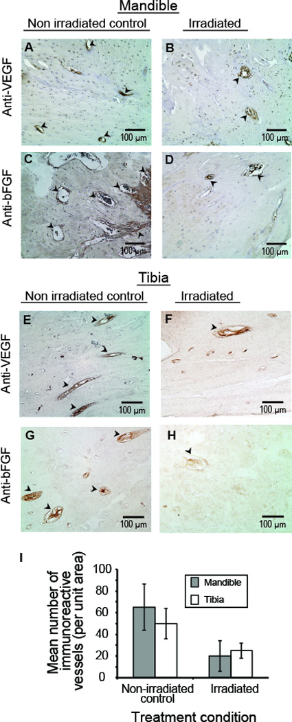
Fig. 6.

Post-irradiation immunoreactive vascular elements. Representative blood vessels immunoreactive to anti-VEGF (A, B, E, F) and anti-bFGF (C, D, G, H) showed a reduction in the number of blood vessels per unit area (black arrowheads) in both irradiated mandible (B, D) and tibia (F, G). Semi-quantitative analysis of immunoreactive vessels (I) showed moderate but non-significant differences between mandible and tibia.
