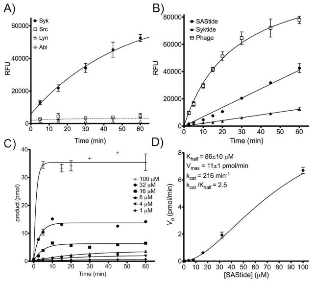
Figure 2.

Phosphorylation of the artificial peptide substrate for Syk (SAStide) in vitro. A) The SAStide biosensor (25 μM) was incubated with Syk-EGFP (closed circles), Lyn, Abl or Src in an in vitro kinase assay. Aliquots were removed at designated time points and the amount of phosphorylated substrate was measured using ELISA-based detection (given as relative fluorescence units, RFU). B) SAStide (GGDEEDYEEPDEPGGKbGG) and two known Syk peptide substrates, ‘Syktide’13a (GGEDDEYEEVGGKbGG) and ‘Phage,’13d a peptide derived from a phage display library (GGEDPDYEWPSAGGKbGG), were incubated with immobilized, immunoprecipitated Syk-EGFP kinase (Syk-EGFP) as described in the Materials and Methods. Substrate concentration for each was 4 μM. C) SAStide was assayed with Syk-EGFP at a range of concentrations. D) Initial velocities were calculated from the data in (C) and plotted and fitted to a sigmoidal curve to fit Khalf, Vmax, kcat and kcat/Khalf. For all panels, data points represent the average of three replicate experiments and error bars indicate the standard error of the mean.
