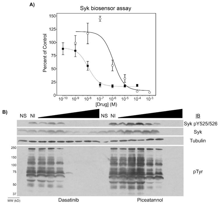
Figure 4.

Detection of Syk inhibitor dose-response. Burkitt’s lymphoma DG75 B cells were treated with varying concentrations of dasatinib (closed squares) or piceatannol (open circles) for 30 min and with the SAStide biosensor (25 μM) for 15 min prior to stimulation. The cells were then stimulated with anti-IgM (5 μg/mL) and hydrogen peroxide (1 mM) for 5 min and harvested. (A) The extent of biosensor phosphorylation was analyzed by ELISA. Data indicate averages +/− SEM of experiments performed in triplicate. (B) The level of total Syk, of Syk phosphorylated on Y525 and Y526 (Syk pY525/526) and of tyrosine-phosphorylated proteins (pTyr) were analyzed by Western blotting of lysates of cells not stimulated (NS), stimulated but not treated with inhibitor (NI) or treated with increasing concentrations of the indicated inhibitor. Tubulin was measured as an internal loading control.
