
Figure 6.
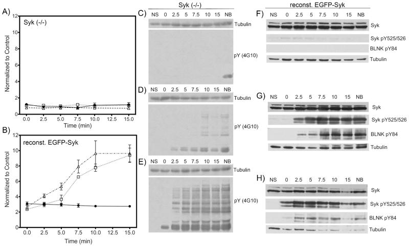
Intracellular specificity of the artificial peptide substrate for Syk. Syk(−/−) DT40 cells or Syk(−/−) DT40 cells reconstituted with stably expressed Syk-EGFP were treated with the SAStide biosensor (25 μM) for 15 min prior to stimulation. The cells were stimulated by treating with anti-IgM (5 μg/mL) (panels C and F, closed circles in graphs A and B), H2O2 (3 mM) (panels D and G, open squares in graphs A and B) or both (5 μg/mL anti-IgM and 3 mM H2O2) (panels E and H, open triangles in graphs A and B), then harvested at varying time points. (C–H): the phosphorylation of proteins in cell lysates was measured by Western blotting using anti-phosphotyrosine (4G10), anti-Syk, antiphospho-Syk(Y525/526), antiphospho-BLNK(Y84) and anti-tubulin antibodies (loading control). NS - no stimulation; NB - no biosensor (15 min harvest). (A, B): chemifluorescence detection of Syk biosensor phosphorylation. The data are reported as normalized change compared to the unstimulated control). Data were analyzed using a repeat measure one-way ANOVA test and a Dunnet post-test; In graph A, no statistically significant difference was seen for any time point or treatment relative to control. Experiments were performed in triplicate.