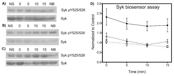
Figure 7.

Detection of Syk activity and inhibition in mouse primary splenic B-cells. Mouse primary splenic B-cells were treated without (panel A and closed circles in panel D) or with the tyrosine kinase inhibitors dasatinib (100 nM) (B), (D △) or piceatannol (50 μM) (C), (D □) for 30 min prior to stimulation, then treated with the SAStide biosensor (25 μM, 15 min) and stimulated with anti-IgM F(ab′)2 (5 μg/μL). Cells were harvested at various time points following stimulation. (A–C) the expression of Syk and of Syk phosphorylated on Y525 and Y526 (Syk pY525/526) were determined by Western blotting. NS - no stimulation; NB - no biosensor (15 min harvest). (D) The data are reported as normalized change in chemifluorescence signal compared to the unstimulated control. N = 6; error bars indicate SEM.
