
Fig. 6. DI-II/gL, DI-II-III/gL and sgH/gL expressed endogenously inhibits cell-cell fusion.
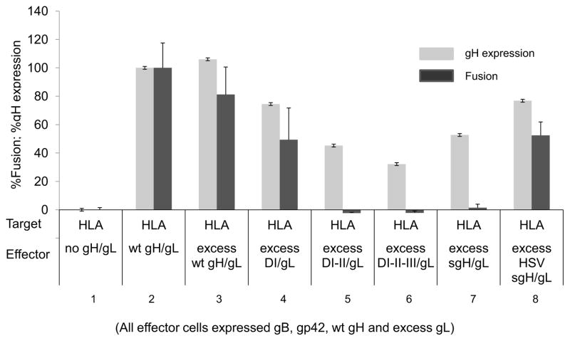
CHO-K1 target cells were transiently co-transfected with HLA-DR and T7 polymerase. CHO-K1 effector cells were transiently co-transfected with gp42, gB, gH, excess gL and either excess wild type gH, HSV gH, DI/gL, DI-II/gL, DI-II-III/gL or sgH/gL. Cells were detached twenty four hours post-transfection and effector cells were overlaid onto target cells. Fusion activity (dark gray bars) was accessed eighteen to twenty four hours post overlay by the addition of passive lysis buffer followed by the addition of luciferase substrate. Luciferase activity was measured with a Perkin-Elmer Victor plate reader. gH/gL expression (light gray bars) was determined by cELISA using anti-gH antibody E1D1. Data shown are representative results of three independent experiments. Error bars represent standard deviations for the normalized values.