
Figure 5. Conditioned running potentiated hippocampal neurogenesis in aging rats.
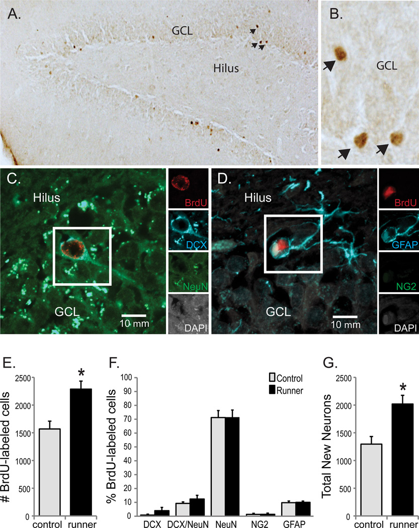
(A) Transmitted light micrograph showing new (BrdU+; in brown) cells located through the GCL and SGZ of aged rats under a 10X objective revealed by DAB. (B) Shows a subset of BrdU+ cells depicted in (A) under the 40X objective used for counting. (C and D) Confocal images of samples of new cells (BrdU/DAPI+; in white and red) expressing the neuronal proteins DCX (in blue) and NeuN (in green; [C]) or the astrocyte protein GFAP (in blue; [D]). Insets show each channel independently and scale bars represent 10 µm. (A, B and E) More BrdU+ cells were detected in the dentate gyri of conditioned runners vs. controls (*p < 0.01). (C, D and F) Similar percentages of new cells in the dentate gyri of conditioned runners and controls expressed immature neuronal (DCX+), transitioning neuronal (DCX/NeuN+), mature neuronal (NeuN+), oligodendroglial (NG2+) or astroglial (GFAP+) proteins. Consistent with the ~2 week survival period, most new cells expressed mature neuronal phenotypes, followed by astrocyte and transitioning neuronal phenotypes. (G) The total estimated new neuron number was significantly higher in conditioned runners versus controls (*p < 0.01). Data are group means ±S.E.M obtained from conditioned runners (black bars) and controls (gray bars).