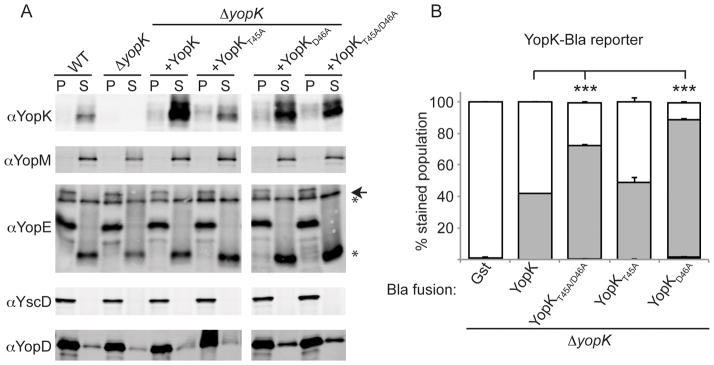
Figure 4. Characterization of YopK point mutants.

(A) ΔyopK Y. pestis carrying a YopK or YopK point mutant expression vector were induced to secrete using temperature switch to 37°C and low Ca2+. Separated by centrifugation, supernatants (S) contain secreted Yops while pellets (P) contain bacteria. YscD is a structural protein of the injectisome and a fractionation control. (arrowhead denotes full length protein while * indicates degradation products) (B) ΔyopK Y. pestis carrying YopK or YopK point mutants fused to Bla were used to infect CHO cells as in panel A. Triplicate samples were averaged and standard deviation is shown. ANOVA followed by Dunnett post-hoc test was done using the ΔyopK +native YopK infection as the control, and *** indicates P<0.001.
