
Figure 8. YopK works within host cells and bacteria.
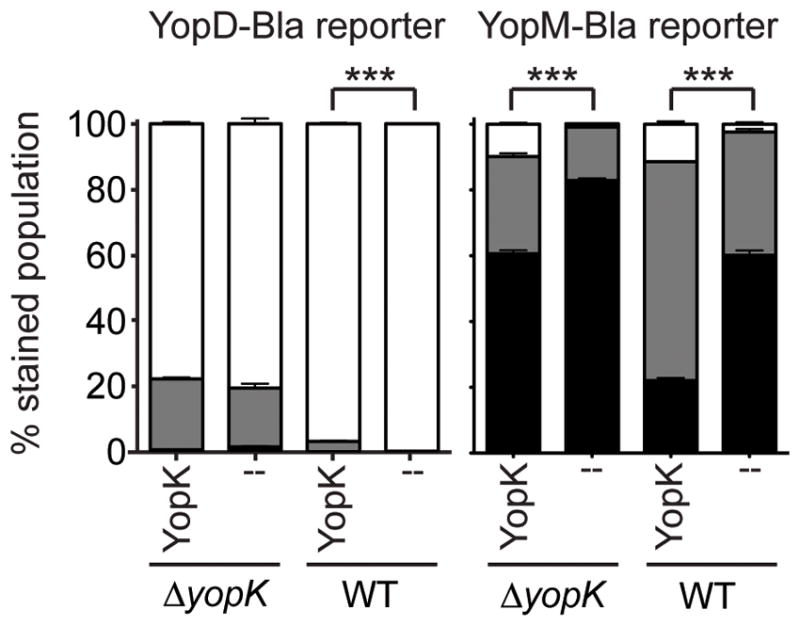
CHO cells were transfected and allowed to express either Keima (--) or Keima-YopK (YopK) for ~36 hours, followed by infection at MOI 10 for 4 hours with Y. pestis strains expressing either YopD-Bla or YopM-Bla. Infected cells were then stained with CCF2-AM and analyzed by flow cytometry. Each infection was performed in triplicate, samples were averaged, and standard deviation shown. Unpaired two-tailed t-tests were done and *** indicates P<0.001. White bars: green cells (uninjected), grey bars: aqua cells (low-level injection), black bars: blue cells (high-level injection).