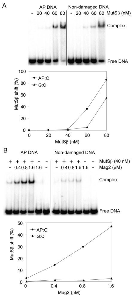
Figure 8. Electrophoretic Mobility Shift Assay With Mag2 and Human MutSβ.

(A) Duplex DNA containing a single AP site or non-damaged DNA (5 nM) was incubated with increasing concentrations of MutSβ (20–80 nM) in a buffer containing 20 mM HEPES, pH 7.5, 2 mM MgCl2, 100 mM NaCl, 5% glycerol, 0.1 mg/ml BSA and 1 mM DTT. Formation of a MutSβ-DNA complex was observed, both for AP DNA and undamaged DNA. (B) Specific binding of MutSβ to AP DNA was stimulated >10 fold by increasing the concentration of Mag2 (0.4–1.6 μM). The amounts of free DNA and DNA in complex with MutSβ were analysed on 10% polyacrylamide gels, visualised using PhosphoImager and quantified using ImageQuant. These data represent a typical result of three independent experiments. See also Figure S4.
