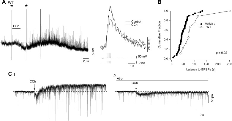
Fig. 10.

Recordings from LDT neurons obtained from WT slices reveal that CCh can induce an inward current and barrage of EPSPs in cholinergic neurons. A: current clamp recording from a cell in the LDT that responded to bath-applied CCh with an initial hyperpolarization and a decrease in the SpECT (right). This hyperpolarization was followed by depolarization and a barrage of EPSP activity. Asterisks (left) mark the 8-Hz train of five spikes producing the SpECTs shown at higher temporal resolution to the right. B: cumulative distribution of excitatory postsynaptic potential (EPSP) barrage latency recorded in slices from M2M4−/− and WT mice. The barrage latency was shifted to longer times in WT mice. C: voltage clamp recordings from an identified LDT cholinergic neuron from a WT slice following puffer application of CCh before (C1) and after the application of atropine (C2). CCh evoked an increase in EPSC frequency and an outward current that were blocked following application of atropine. Note the early atropine-resistant inward current which is attributable to activation of nicotinic acetylcholine receptors by the rapid application method.
