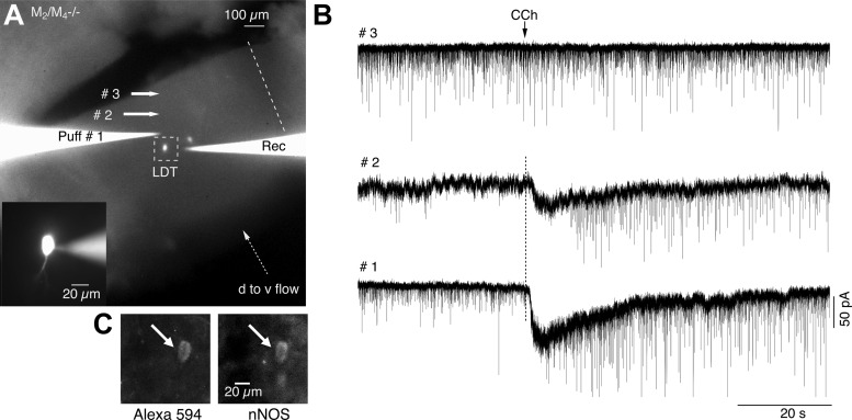
Fig. 9.

CCh activates local glutamatergic neurons. A: low power fluorescence image of the LDT which has been surgically isolated from more ventral portions of the slice. Midline is indicated by the dotted line, and the dorsal-to-ventral flow of ACSF is indicated by the dotted arrow (d to v flow). Both the recording pipette (right) and puff pipette (Puff #1) contain Alexa 594 and are fluorescent, as are two recorded and filled neurons which are visible. Inset shows the leftmost neuron at higher magnification during the recording (pipette seen on right side). Responses shown in B were obtained from this neuron, with the puff pipette located at the corresponding positions. B: the increase in sEPSC frequency and postsynaptic inward current decreased as the puff location was moved out of the LDT. C: immunocytochemistry following fixation revealed the recorded cell was nNOS+ and hence cholinergic.
